Study reveals why the brain ‘zones out’ when you’re exhausted

We’ve all experienced the intense difficulty of staying attentive after a bad night’s sleep – and a new study shows what happens in the brain when this feeling arises.
When you walk away after a sleepless night, the brain flushes out cerebrospinal fluid (CSF), which surrounds the brain and spinal cord and is one of the waste disposal system. This CSF then returns to the brain when you exit it, according to the study published October 29 in the journal Natural neuroscience.
“By measuring so many different types of information about the brain simultaneously, we were able to see that these many different things that we initially thought were separate were actually moving together,” co-author of the study. Laura Lewisassociate professor of neuroscience at MIT, told Live Science.
Sleep is critical for maintain a healthy brainand experts recommend adults seven to nine hours of sleep one night. Not getting enough sleep takes a toll on a person’s mental and physical health and also impairs our ability to pay attention. “However, the neural basis of attentional failures induced by sleep deprivation is not yet well understood,” the authors write in the study.
To investigate, they recruited 26 healthy volunteers aged 19 to 40, including 19 women. All participants participated in two study conditions approximately 10 days apart: well-rested and sleep-deprived. Half finished the session well-rested first, while the others started with a lack of sleep.
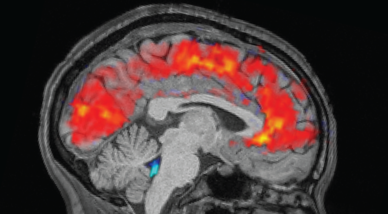
Rested individuals slept between 6.5 and 9 hours at home, while sleep-deprived individuals stayed awake all night in the laboratory. The morning before each trial, the team attached electroencephalogram (EEG) caps to participants to record their brain waves with electrodes. Simultaneously, participants underwent functional testing MRI (fMRI) to reveal patterns of blood and CSF flow in the brain. Eye trackers measured the size of the participants’ pupils.
These are things we don’t typically think of as being closely related to time: your ability to pay attention to the world and then to basic fluid movements in the brain.
Laura Lewis
Participants then completed tasks that required their visual and auditory attention; they pressed a button as soon as they saw an image or heard a noise. Additionally, the researchers collected data on baseline brain activity by asking participants to rest for 25 minutes without performing any tasks.
As expected, participants took longer to notice the stimuli when they were tired and missed the cues more often than when they were well rested. But the team was surprised to see giant CSF pulses in exhausted individuals, alongside slow brain wave patterns – both of which are normally seen in exhausted individuals. non-REM sleep.
Specifically, the patterns resembled those observed when a person moves from step N1 to N2the first two of three stages of non-REM sleep that people experience every sleep. “This was something we had only seen before on this scale during sleep,” Lewis said.
CSF flow was closely related to pupil size, with large inflow following pupil dilation and outflow following pupil constriction. This link was more pronounced in sleep-deprived individuals, which could suggest that the body’s circulatory system underlies this coupling, the authors wrote. CSF flows also coincided with when individuals moved during the tasks.
“When you have attention problems… this fluid is drained from your brain, and when you regain your attention, when you start responding to stimuli again, this fluid returns to the brain,” explains the first author of the study. Zinong Yanga computational neuroscientist at MIT, told Live Science.
“These are things that we don’t typically think of as being closely related to time: your ability to pay attention to the world and then to basic fluid movements in the brain,” Lewis added.
The researchers believe that the brain patterns they observe may reflect the fact that the brain, deprived of sleep, enters a sleep-like state, but while still awake. Losses of attention signal the start of these sleep-like brain processes, but they are interrupted before sleep properly takes hold.
But for now, the functional reason behind the huge changes in blood flow is unclear, Lewis noted. Future work could examine whether and how these patterns affect the removal of toxic metabolic waste from the brain, the authors wrote.
Michael Cheedirector of the Center for Sleep and Cognition at the National University of Singapore, who was not involved in the study, said the research was “an impressive piece of physiology.” He thinks that autonomy nervous systemwhich controls unconscious bodily functions, drives these signals.
“I think the big lesson is, ‘Hey, listen; this humble autonomic system that we don’t really pay attention to actually orchestrates some of the biggest signal changes, pupil changes, EEG signal changes, BOLD signals. [fMRI] changes,” he told Live Science.
However, Chee pointed out that participants experienced 24 hours of sleep deprivation, while most people only lose a few hours of sleep on a bad night. “It’s a heavy manipulation,” he said, so “these are disproportionate effects.”
Studying these brain changes in people with sleep problems could point to new therapeutic targets, Chee added.


