Human Embryo Implantation Revealed in First-Ever 3D Images

August 15, 2025
3 Min read
The first 3D images of the establishment of human embryos reveal new details of the process
Analysis of embryo movements in environments in a uterus type could offer indices to improve the success rate of in vitro fertilization
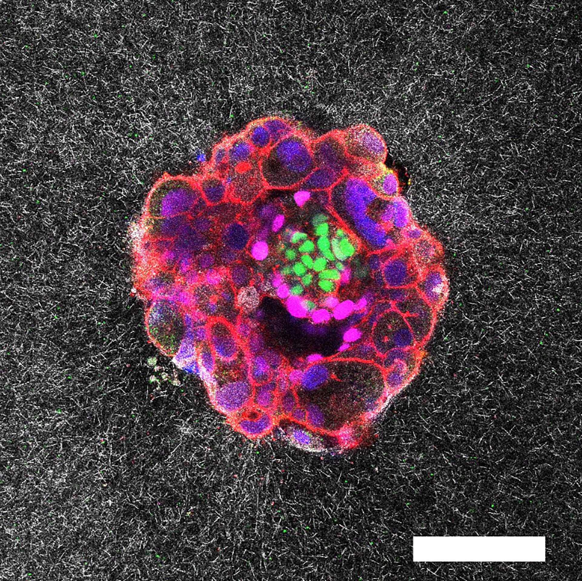
Image of confocal microscopy of a nine -day human embryo. Specific proteins and cellular structures have been colored in the image: Oct4 (green), which is linked to embryonic stem cells; Gata6 (Magenta), which is associated with early tissue training; DAPI (Blue), which marks DNA in nuclei; and Phalloidine (red), which reveals the actin cytoskeleton. The scale bar corresponds to 100 µm.
Catalonia Bio-Engineering Institute (IBEC)
The researchers captured the first three -dimensional images in real time and three -dimensional of a human embryo set up in collagen designed to imitate uterine tissues – a key stage of reproduction. The resulting images, which show how embryos grow and pull to anchor in the uterus in detail, could lead to improvements in in vitro fertilization techniques (IVF), according to scientists.
“This will allow us to develop treatments specifically targeting the establishment, which is the largest road dam in human reproduction,” explains Samuel Ojosnegros, bio-engineer at the Barcelona Institute of Science and Technology in Spain and co-author of the new study, which was published in Scientific advances.
Five days after an embryo is artificially fertilized, fertility doctors must implant it in the body so that he can continue to grow. “What is happening between the transfer and the first ultrasound of weeks later is a black box,” explains Ojosnegros, who is also co-founder of the Biotech Company Serabiotics. The failure of the implantation is one of the main causes of infertility – up to 60% of false layers occur during this process.
On the support of scientific journalism
If you appreciate this article, plan to support our award -winning journalism by subscription. By buying a subscription, you help to ensure the future of striking stories about discoveries and ideas that shape our world today.
The first successful culture of human embryos beyond the establishment was demonstrated in a petri box in a laboratory in 2016, but Ojosnegros and his team wanted to see what this process would look like in the 3D fabric which was more similar to that of the uterus.
To do this, the team designed a special ex vivo system in frost and collagen – a protein found in the uterine lining – and used embryos given by people who had finished an assisted reproductive process. The system works, says Ojosnegros, because the collagen fiber network signals to the embryo at a molecular level that it is a natural matrix.
Using advanced 3D microscopes, the researchers recorded the action over time. The follow -up of tiny movements in the frost of the frost allowed them to map exactly where and how much the embryos drew. The researchers did the same with mouse embryos to compare movement models.
Images have shown that human embryos generate a network of tiny traction forces in the uterus. They dig into the surrounding fabrics on one side, creating several small traction points that draw the lining in all directions. Mouse embryos, on the other hand, spread more on the surface and draw mainly along two or three strong lines.
Compacting embryo and invading uterine fabric.
When the researchers applied an external tension to the matrix, pulling it with tiny pliers, they noticed that the embryos reoriented themselves to these areas. Scientists suggest that micro-contracts could guide the embryo to the implant in the optimal direction of the uterus. “We believe that these micro-contracts are what the embryo uses to guide itself to the blood vessels and the nutrients it needs,” explains Ojosnegros, adding that more studies are necessary to confirm this hypothesis.
In mouse and human experiences, the strength and scheme of these forces were linked to the health of the embryo, which means that the embryos that fired less were less likely to invade the fabric. The observation of real -time implantation in a 3D model is a “quantum jump” compared to the two -dimensional observations which already exist, explains that the biologist of development Claudia Spits of the Free University of Brussels, which was not involved in research. Keeping an embryo alive under these conditions is extremely difficult, she said. “What you see in a 10 -second video is years to define these [conditions] So that the embryo can survive, ”adds Spits.
Two embryos implanting in the uterus.
“This study opens the way to the exploration of the dynamics of implantation in unprecedented details,” explains Magdalena & Zdot; Ernicka-Goetz, development biologist at the California Institute of Technology, who was not involved in research. The results are added to all the work on human post-implantation observations published in the past nine years, she says, and “these studies are a step forward in the understanding of a stage of human development which has long been hidden from vision”. Future research notes, & zdot; Ernicka-Goetz are still necessary to compare how embryos behave on different platforms “similar to a uterus” to see if the development trajectories differ.
The matrix developed by the Ojosnegros team is not intended for in vitro fertilization procedures, but it could be a precious tool for pharmaceutical companies and laboratories testing serums or different types of embryos. “By starting to understand how the embryo behaves,” says Ojosnegros, “we can start thinking about the future possibility of selecting healthy embryos or those more capable of implanting.” Pins remain skeptical about this assertion because the replication of this technology in other laboratories could be a major challenge. But she says that the results are a “big advance” in technology that could have future applications once other laboratories are able to make their own 3D locations.


