Science history: Doctor autopsies the brain of a man who couldn’t speak — and reveals the seat of spoken language — April 18, 1861

QUICK FACTS
Milestone: Autopsy of the famous patient “Tan”
Date: April 18, 1861
Or: Bicêtre Hospital, Paris region
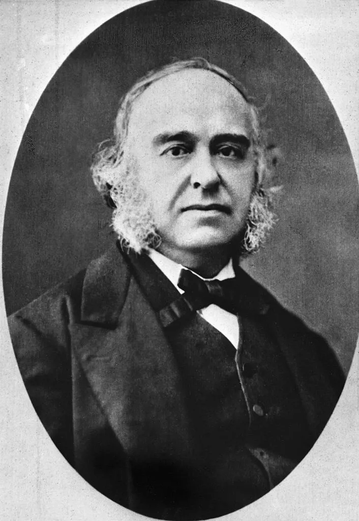
WHO: Dr Paul Broca
On April 18, 1861, a Parisian doctor opened the brain of a patient who had died the day before and unwittingly identified a brain region essential for spoken language.
The patient, Louis Victor Leborgne, was nicknamed “Tan” by the doctors at Bicêtre hospital because it was one of the only words he could pronounce. At the time of his death at the age of 51, he had spent 21 years in the hospital’s psychiatric ward.
Leborgne was reportedly healthy at birth, but began having epileptic seizures in early childhood. At age 30, he lost the ability to speak. For a time he avoided seeking treatment, but was eventually admitted to Bicêtre hospital.
Article continues below
Doctors found that he understood language well and used gestures to express his needs. Rarely, he could say a swear word.
A decade after his admission to the hospital, he began suffering from progressively worsening right-sided paralysis, as well as mental disorders. Eventually, he lost the ability to walk. He spent the last seven years of his life in bed.
In recent years, Dr. Paul Broca, a surgeon at the hospital, began to treat Leborgne as a patient.
“Numerical responses were the ones he did best, by opening or closing his fingers. He indicated, without error, the time to the nearest second on a watch. He knew exactly how many years he had been in Bicêtre, etc.” Broca said of his patientaccording to one translation.
“However, many questions that a man of normal intelligence would have found a way to answer with gestures remained without an intelligible answer; other times the answer was clear, but did not answer the question,” observed Broca. “Undoubtedly the patient’s intelligence had therefore been affected to a great extent, but he certainly retained more than was necessary for speech.”
On April 17, 1861, Leborgne died of gangrene, probably due to a bedsore on his leg. The next day, Broca began an autopsy and noticed a pocket of clear fluid the size of a “chicken egg” in the perisylvian region of the left hemisphere of the brain; this region surrounds a deep furrow called the lateral furrow, which marks the upper limit of the temporal lobe. Several areas surrounding the fluid exhibited “smoothness.” And there were other abnormalities: Leborgne’s brain was lighter than normal and several brain regions had a smaller volume than expected.
The same day, Broca presented the results of his autopsy at the Meetings of the Anthropological Society in Paris. At the time, a debate raged between scientists who believed that all of the brain’s functions were distributed throughout the organ’s tissues and those who believed that certain regions performed specific functions.
Broca’s autopsy was strong evidence for the latter idea.
“The main focus and original site of flabbiness is the middle part of the frontal lobe of the left hemisphere; this is where the most extensive, most advanced and oldest lesions are found,” he said in his presentation.
This suggests that “in the present case, the frontal lobe lesion was the cause of the speech loss,” Broca added.
At the meeting, however, his peers did not immediately recognize the significance of this discovery; most of the meeting was devoted to now discredited discussions Racial “science” has focused on supposed links between skull measurements and intelligence. But by August 1861, Broca had studied the brains of several patients suffering from what would later be called aphasia. The research reinforced his belief that speech was localized in the frontal lobeand it will later shrink the region to the left frontal lobe.
During his life, Broca not only identified the region linked to aphasia but also noted that speech therapy could occasionally help patients regain speech.
Since Broca’s time, researchers have confirmed that distinct brain regions perform specific cognitive functions and have focused on a much more precise brain region that is essential for speech than that identified by Broca. This area is now called the Broca district and is recognized as being important in Broca’s aphasia, in which patients can understand language but have difficulty producing spoken, written, or sign language.
We now know that other regions and networks beyond the Broca area play a big role in the speech. For example, lesions of Wernicke’s area, discovered in 1874, can trigger a form of aphasia in which patients speak in long, complete sentences that make little sense.
For decades, Leborgne’s intact brain, which Broca never cut into sections but only superficially examined, could be observed in daylight. Dupuytren Museum in Parisclosed to the public in 2016.


